 ultimate-guide
Long-Term Effects of Untreated Meibomian Gland Dysfunction
Table of Contents
- What Is Meibomian Gland Dysfunction and Why It Matters
- How Meibomian Gland Dysfunction Affects the Tear Film and Ocular Surface
- Meibomian Gland Dysfunction Prognosis: What Happens Without Treatment
- Untreated Dry Eye Complications: Corneal Damage and Vision Loss
- Gland Atrophy and Permanent Loss: Can the Damage Be Reversed?
- Home Treatments for Meibomian Gland Dysfunction and Daily Management
- How Long Does Meibomian Gland Expression Last and When to Seek Professional Care
- Conclusion: Managing MGD Before Complications Develop
- Frequently Asked Questions
Last Updated: October 4, 2026
What Is Meibomian Gland Dysfunction and Why It Matters
Meibomian gland dysfunction is a chronic condition in which the oil-producing glands along the eyelid margins fail to secrete adequate meibum, the lipid layer essential for tear-film stability. This disruption creates a cascade of ocular surface problems that, left untreated, can progress from mild discomfort to permanent structural damage. (Source: National Eye Institute's information on dry eye disease)
When meibomian glands become obstructed or atrophy, the tear film loses its protective oil component, allowing tears to evaporate rapidly. Many patients dismiss early symptoms as temporary irritation, only to discover years later that significant gland damage has already occurred.
Meibomian gland dysfunction progresses slowly. Early symptoms, mild dryness, grittiness, or blurred vision, seem manageable, but without intervention, glands atrophy permanently, making reversal increasingly difficult.
Early detection and management halt progression and preserve gland function. Delayed treatment risks chronic discomfort and vision-threatening complications.
How Meibomian Gland Dysfunction Affects the Tear Film and Ocular Surface
The tear film's outer lipid layer, produced by meibomian glands, prevents tear evaporation and maintains stability. When meibomian gland dysfunction disrupts this layer, consequences ripple across the entire ocular surface.
Without adequate meibum, tears evaporate rapidly, leading to tear hyperosmolarity. The cornea and conjunctiva respond with inflammation, damaging epithelial cells and further reducing tear quality in a vicious cycle.
Epithelial cells become damaged, the conjunctiva reddens, and patients experience burning, grittiness, and fluctuating vision. This evaporative dry eye disease is the direct result of meibomian gland dysfunction.
This inflammation is progressive; each cycle damages more cells and sets the stage for severe complications.
Meibomian Gland Dysfunction Prognosis: What Happens Without Treatment
Untreated meibomian gland dysfunction follows a predictable but variable pattern. Understanding the timeline and likelihood of outcomes is essential for realistic planning.
Stage 1: Early Obstruction (Months 1-12)
In early obstruction, glandular tissue remains structurally intact. Symptoms are mild: intermittent dryness, grittiness, or blurred vision worsening toward evening or after screen use.
Early obstruction is highly reversible. Glands obstructed for weeks to a few months respond well to warm compresses, eyelid hygiene, and professional expression.
Stage 2: Progressive Obstruction and Early Gland Loss (Year 1-3)
Without treatment, obstruction spreads and triggers gland dropout. Meibography reveals gaps where healthy glands once were.
Gland loss varies: some patients lose 20-30% over 2-3 years; others lose 40-50%.
Partial recovery is possible with aggressive treatment: frequent professional expression, strict daily hygiene, and possible oral medications may stabilize function or recover 40-70% of lost capacity. Without intervention, progression accelerates.
Stage 3: Advanced Atrophy and Permanent Loss (Year 3+)
After 3+ years unchecked, atrophied glands undergo fibrosis and damage becomes irreversible. No treatment regenerates lost glands. Meibography shows 60-80%+ gland dropout.
Symptoms are severe: constant dryness, blurred vision, photophobia, foreign-body sensation, and pain affecting daily function. Patients struggle with reading, driving, and screen work. Sleep is disrupted by nocturnal dryness.
Approximately 10-15% develop clinically significant corneal involvement: punctate erosions, filamentary keratitis, or corneal ulceration or scarring. These are largely preventable with earlier intervention.
Variability and Individual Risk Factors
Not all patients follow this timeline identically. Age, sex, genetics, and systemic health significantly influence progression rate:
Patients over 50 progress faster. Menopause and androgen deficiency accelerate loss. Antihistamines, isotretinoin, SSRIs, and anticholinergics impair secretion. Poor contact lens hygiene increases dropout risk. Rosacea and Sjögren's accelerate progression. Daily home care stabilizes function; neglect accelerates decline.
The Critical Window
The first 12-24 months represent a critical window. Early detection and prompt treatment preserve gland function and prevent complications.
Untreated Dry Eye Complications: Corneal Damage and Vision Loss
Untreated meibomian gland dysfunction causes corneal damage and functional impairment. Both are largely preventable with early intervention.
Corneal and Ocular-Surface Damage
As meibomian gland dysfunction progresses, the cornea faces hyperosmolar tears, reduced tear volume, chronic inflammation, and mechanical friction from unstable blinking.
Vital dyes reveal punctate erosions, small areas of missing epithelial cells, representing actual cell loss from dry conditions and inflammatory mediators.
Persistent dryness deepens erosions. Filamentary keratitis develops, causing severe blurred vision, fluctuation, and substantial pain requiring urgent intervention.
Corneal ulceration, full-thickness epithelial loss, can progress to scarring or perforation. Scarring causes permanent vision loss; perforation is a medical emergency.
Functional and Quality-of-Life Complications
Vision fluctuation: Clarity changes moment to moment as the tear film becomes unstable.
Screen intolerance: Incomplete blinking during screen use reduces tear-film refresh. Patients struggle with concentration, take frequent breaks, and reduce work hours.
Reading difficulty: Sustained near focus worsens symptoms. Some abandon reading for pleasure, substantially affecting quality of life. (Source: American Academy of Ophthalmology's guidelines on meibomian gland dysfunction)
Driving safety: Fluctuating vision, glare sensitivity, and photophobia impair safety, especially at night. Some restrict driving to daytime; older patients may stop driving, losing independence.
Sleep disruption: Nocturnal dryness causes patients to wake with severe discomfort. Sleep quality suffers, leading to daytime fatigue and reduced cognitive function.
Prevalence and Severity of Complications
Complication likelihood depends on disease stage, duration, and individual factors:
Early stage (<2 years): 80-90% have normal corneal appearance. Moderate-to-advanced (2-5 years): 30-40% develop corneal staining. Advanced (>5 years): 10-15% develop significant corneal involvement with severe functional limitation.
Why Early Intervention Matters
Early intervention (within 6-12 months) avoids corneal damage and functional limitation. Late intervention (after 3-5 years) risks chronic impairment and complications.
Gland Atrophy and Permanent Loss: Can the Damage Be Reversed?
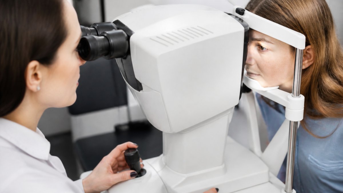
Reversibility depends on treatment stage. Early obstruction is highly reversible with warm compresses, eyelid hygiene, and professional expression, restoring function in weeks to months.
Persistent obstruction causes atrophy, partial glandular loss. Remaining tissue retains some treatment response. Patients with moderate atrophy may recover partial function; complete recovery is unlikely but stabilization is achievable.
Permanent gland loss occurs when atrophied glands undergo complete fibrosis. The glandular tissue is replaced by scar tissue. At this stage, the damage is irreversible. No treatment can regenerate lost glands or restore their function. The goal shifts from recovery to management and slowing further decline.
The distinction matters clinically. A patient with early atrophy might recover 60-80% of function with intensive treatment. A patient with advanced atrophy might stabilize at 20-30% function. A patient with permanent gland loss cannot recover function but can manage symptoms to prevent further damage.
This is why imaging matters. Meibography allows us to quantify gland loss and stage the disease. It informs prognosis and guides treatment intensity. A patient with 50% gland dropout is in a different situation than one with 80% dropout, even if symptoms are similar.
Home Treatments for Meibomian Gland Dysfunction and Daily Management
Management of meibomian gland dysfunction begins at home. Consistent daily practices can slow progression, reduce symptoms, and in early stages, reverse obstruction entirely.
Warm compresses are the foundation. Heat liquefies meibum, making it flow more freely from the glands. Apply a warm compress (around 110°F) to closed eyelids for 10-15 minutes daily, ideally in the morning.
Eyelid hygiene follows immediately after warmth. Gently massage the eyelid margins in a rolling motion toward the lash line, using light pressure. This mechanical action helps express meibum from the glands.
Lid hygiene products designed for meibomian gland dysfunction, such as eyelid cleansers formulated to remove biofilm and bacterial overgrowth, can improve outcomes. These products address the chronic low-grade inflammation that perpetuates obstruction.
Omega-3 supplementation has evidence supporting its use. Omega-3 fatty acids improve tear composition and reduce inflammation. Typical doses are 1,000-2,000 mg daily of combined EPA and DHA. Effects develop over weeks to months, requiring patience and consistency.
Environmental modifications matter. Reduce screen time or implement the 20-20-20 rule: every 20 minutes, look at something 20 feet away for 20 seconds. Blinking deliberately during screen use helps distribute meibum across the ocular surface.
Artificial tears provide symptomatic relief but don't address the underlying problem. Lipid-based tears mimic the natural tear film more closely than aqueous tears alone and are preferable for meibomian gland dysfunction.
How Long Does Meibomian Gland Expression Last and When to Seek Professional Care
Professional meibomian gland expression, performed by an eye care provider using specialized equipment and technique, is more effective than home expression alone. The provider applies controlled heat and mechanical pressure to express meibum from obstructed glands.
The effects of a single expression session typically last 2-4 weeks. Some patients experience symptom relief immediately; others notice improvement over several days as inflammation subsides. The duration varies based on disease severity, gland obstruction density, and individual healing response.
For patients with moderate meibomian gland dysfunction, professional expression every 4-6 weeks, combined with daily home care, can maintain gland function and prevent progression. For severe cases with extensive obstruction, more frequent sessions, every 2-3 weeks, may be necessary.
The key insight is that expression is not curative; it's maintenance. It clears existing obstruction but doesn't prevent new obstruction from forming. Without ongoing home care and periodic professional treatment, glands reobstruct within weeks.
Seek professional care immediately if you experience:
- Persistent eye pain or severe discomfort
- Sudden vision changes or blurred vision that doesn't resolve with blinking
- Eye redness accompanied by discharge or crusting
- Photophobia (light sensitivity) that interferes with daily function
- Signs of corneal involvement such as foreign-body sensation that worsens despite drops
Additionally, if home care fails to improve symptoms after 4-6 weeks of consistent practice, professional evaluation is warranted. At Wilck Schwartz & Novak OD PC, we use advanced optometric equipment to assess gland structure, quantify gland loss, and determine whether your condition requires escalation to more intensive treatment.
Regular monitoring, annual or biannual eye exams, allows us to track meibomian gland dysfunction progression before symptoms become severe. Early detection of gland atrophy enables intervention while recovery is still possible.
Conclusion: Managing MGD Before Complications Develop
Meibomian gland dysfunction is progressive, but it is not inevitable. The difference between a patient who manages this condition successfully and one who develops corneal scarring or permanent vision loss often comes down to timing: how early the condition was detected and how consistently it was treated.
The challenge is that early meibomian gland dysfunction feels manageable. Mild dryness and occasional discomfort don't seem urgent. But beneath the surface, glands are obstructing and beginning to atrophy. The window for reversing this process is limited.
At Wilck Schwartz & Novak OD PC, we're committed to early detection and precise treatment of meibomian gland dysfunction. Our advanced optometric equipment allows us to visualize gland structure, assess tear-film quality, and stage disease severity with accuracy that routine eye exams often miss. We diagnose ocular diseases with greater precision and manage them with a focus on long-term outcomes rather than short-term symptom relief.
If you're experiencing persistent dry eye, fluctuating vision, or chronic eye discomfort, schedule a comprehensive eye exam. Early intervention can preserve gland function and prevent the complications that define advanced meibomian gland dysfunction. Your vision and quality of life depend on it.
Frequently Asked Questions
What is the prognosis for meibomian gland dysfunction if left untreated?
Without treatment, meibomian gland dysfunction typically worsens over time. The glands become progressively obstructed, reducing oil secretion and destabilizing the tear film. This leads to increased tear evaporation, corneal surface damage, and chronic discomfort. Some patients experience gland atrophy and permanent loss of function. Early intervention can slow this progression and prevent irreversible damage.
Can untreated meibomian gland dysfunction cause permanent vision loss?
Prolonged, untreated meibomian gland dysfunction can result in corneal damage severe enough to affect vision quality. Corneal staining, scarring, and ulceration are possible complications. While total blindness is rare, chronic corneal inflammation and epithelial damage can cause persistent blurred vision, fluctuating vision, and light sensitivity. Professional diagnosis and management are essential to prevent these outcomes.
How long does meibomian gland expression last as a treatment?
Meibomian gland expression provides temporary relief, typically lasting 2-4 weeks. The benefit duration varies based on gland health, obstruction severity, and individual tear-film chemistry. Regular professional expression, combined with daily warm compresses and eyelid hygiene, helps maintain gland function. For lasting improvement, expression is most effective when paired with consistent home care and professional monitoring.
Can meibomian gland atrophy be reversed with home treatments?
Once gland atrophy occurs, the damage is usually permanent and cannot be fully reversed with home treatments alone. However, early intervention with warm compresses, eyelid hygiene, and professional gland expression can slow atrophy and preserve remaining function. This is why early diagnosis and consistent management are critical. Advanced atrophy may require prescription treatments to manage symptoms and prevent further deterioration.
Don't let untreated meibomian gland dysfunction progress to permanent gland loss. Schedule an appointment with Wilck Schwartz & Novak OD PC today and take control of your ocular health before complications develop.