 ultimate-guide
Advanced Eye Care Technology in Brooklyn
Table of Contents
- What Advanced Eye Care Technology Really Means
- What Is an OCT Eye Scan and Why It Matters
- Digital Retinal Imaging Benefits for Early Detection
- Comprehensive Eye Exams With State-of-the-Art Equipment
- Laser Vision Correction and Advanced Surgical Options
- How to Choose an Eye Doctor With Advanced Technology
- Why Technology Matters for Your Entire Family's Vision
- Frequently Asked Questions
Last Updated: August 12, 2026
What Advanced Eye Care Technology Really Means
Advanced eye care technology refers to modern diagnostic and treatment equipment that detects eye diseases earlier, fits contact lenses more precisely, and corrects vision problems with greater accuracy than traditional methods. At Wilck Schwartz & Novak OD PC, advanced technology integration means patients receive comprehensive evaluations that catch problems most standard eye exams would miss.
Standard vision checks measure whether you see 20/20 and update your glasses prescription. Advanced technology examines deeper eye structures, retina, optic nerve, cornea, and macula, revealing early signs of glaucoma, diabetic retinopathy, macular degeneration, and other sight-threatening conditions before symptoms appear. Vision and eye health are not the same: you can have perfect 20/20 vision and still have glaucoma developing silently, damaging your optic nerve without warning. Advanced eye care technology catches these conditions in their earliest stages, when treatment is most effective.
What Is an OCT Eye Scan and Why It Matters
An OCT eye scan, or optical coherence tomography, is a non-invasive imaging technique that creates detailed cross-sectional images of the retina and optic nerve using light waves to map tissue layers with remarkable precision.
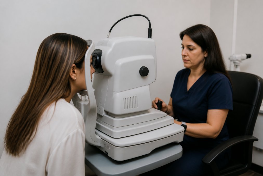
The OCT scan directs near-infrared light into your eye and measures reflection time. A computer constructs a detailed map showing the thickness and structure of each retinal layer. The entire scan takes about 30 seconds per eye and produces images with resolution measured in microns, far more detailed than traditional examination tools. The procedure is non-invasive, requires no contact with your eye, no dilation, and causes no discomfort.
OCT imaging detects glaucoma before you notice any vision loss by identifying early thinning of the nerve fiber layer years before functional vision loss becomes measurable. For patients with diabetes, OCT reveals swelling in the macula before vision becomes blurry, allowing treatment to prevent serious vision loss. For anyone over 55, OCT screens for age-related macular degeneration, the leading cause of vision loss in older adults, at a stage when intervention can slow progression significantly.
Digital Retinal Imaging Benefits for Early Detection
Digital retinal imaging captures high-resolution photographs of the back of your eye, documenting the health of your retina, optic nerve, and blood vessels. Unlike traditional retinal examination, which relies on examiner judgment, digital imaging creates a permanent record for year-to-year comparison to detect subtle changes.
Diabetic retinopathy often shows no symptoms until damage is advanced. Digital imaging reveals the earliest signs, microaneurysms, dot hemorrhages, and hard exudates, that signal disease progression, allowing coordinated treatment to prevent blindness. Hypertensive retinopathy, caused by high blood pressure damaging retinal blood vessels, also appears on digital imaging before causing vision problems, providing information to share with your primary care physician for medication adjustments.
For patients with no systemic disease, digital retinal imaging documents your retina's normal appearance, creating a baseline for comparison. If you develop a new floater, flash of light, or shadow in your vision months or years later, your eye care provider can compare new imaging to your baseline and determine whether the change is benign or requires urgent attention. Modern digital retinal imaging systems capture high-definition details and wide-angle views up to 200 degrees of the retina in a single shot, catching peripheral retinal problems that might otherwise go unnoticed.
Comprehensive Eye Exams With State-of-the-Art Equipment
A comprehensive eye exam with advanced eye care technology extends far beyond basic vision checks to assess the overall health of your ocular structures and visual system.
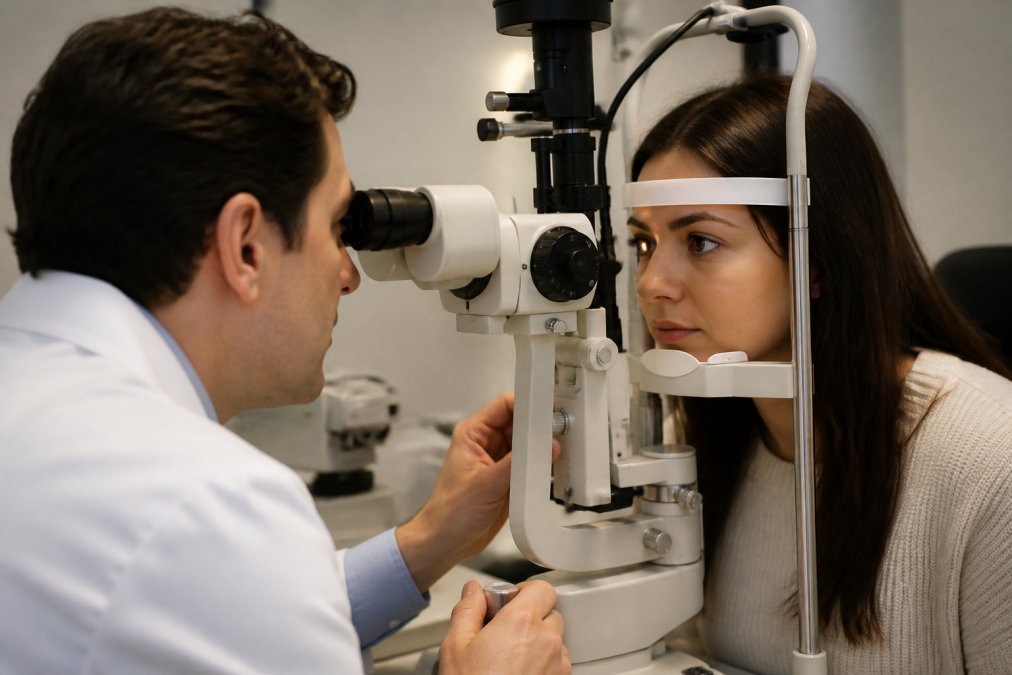
The exam begins with a detailed visual history covering vision changes, eye strain, dry eye symptoms, family history of eye disease, and systemic conditions affecting your eyes. Visual acuity measurement follows using a standard eye chart, but modern comprehensive exams extend further. Corneal topography maps the shape of your cornea with precision, essential for fitting contact lenses properly and detecting astigmatism or keratoconus. Biometry measures eye length, crucial for anyone considering cataract surgery or refractive surgery.
Intraocular pressure testing screens for glaucoma, a critical risk factor requiring monitoring. Pupil dilation, when performed, allows examination of the lens, vitreous, and peripheral retina. Many practices now use non-dilated digital imaging to capture retinal detail, reducing the need for dilation in routine exams.
A comprehensive exam with advanced equipment typically takes 45 minutes to an hour, allowing thorough assessment of your visual system and early detection of problems affecting your sight. For families seeking a single provider for pediatric and adult eye care, comprehensive exams ensure children receive the same level of diagnostic assessment as adults, catching refractive errors and binocular vision problems early in development.
Laser Vision Correction and Advanced Surgical Options
Laser vision correction, commonly known as LASIK or PRK, reshapes the cornea to correct refractive errors, potentially eliminating the need for glasses and contact lenses. Modern systems offer customized treatments based on detailed mapping of each patient's unique corneal shape.
A femtosecond laser creates a thin corneal flap (in LASIK) or the outer layer is gently removed (in PRK). An excimer laser then reshapes the underlying corneal tissue with precision measured in microns. The procedure takes about 15 minutes total, with the actual laser application lasting only 20-40 seconds per eye.
Wavefront-guided LASIK uses detailed mapping of how your eye focuses light, revealing subtle irregularities beyond simple myopia or hyperopia. The laser treatment corrects not just your prescription but also higher-order aberrations, potentially improving your vision beyond 20/20. Topography-guided treatments customize the laser pattern further, particularly beneficial for patients with irregular astigmatism or previous corneal scars.
Advanced diagnostic imaging determines candidacy for laser vision correction. Corneal thickness must be adequate to allow safe reshaping. Corneal topography must show no signs of keratoconus or other ectatic diseases that would contraindicate surgery. Optical coherence tomography assesses anterior segment structures, ensuring the cornea, lens, and other tissues are healthy enough for the procedure.
Cataract surgery has also been transformed by advanced eye care technology. Modern phacoemulsification uses ultrasonic energy to break up the clouded lens, followed by intraocular lens (IOL) implantation. Premium multifocal or extended-range IOLs correct presbyopia, potentially reducing dependence on reading glasses after surgery. Toric IOLs correct astigmatism during cataract surgery, addressing multiple vision problems simultaneously.
Optical biometry uses light-based measurement accurate to within 0.1 mm, dramatically improving IOL power calculations. This precision means more patients achieve their target refraction without needing glasses after cataract surgery.
How to Choose an Eye Doctor With Advanced Technology
Choosing an eye care provider involves more than convenience and insurance coverage. The equipment and technology available directly affects the quality of care you receive and your ability to detect eye disease early.
Ask what diagnostic equipment the practice uses: OCT imaging, digital retinal imaging, corneal topography, and automated visual field testing represent the standard of modern eye care. Inquire about the scope of services offered. Does the practice handle emergency eye care, diagnose and manage ocular diseases, offer contact lens fittings, and evaluate candidates for laser vision correction? A comprehensive practice reduces the need to visit multiple providers.
Ask about the eye care provider's credentials and experience. Optometrists (OD) complete four years of optometry school and are licensed to perform comprehensive eye exams, diagnose many eye diseases, and prescribe medications. Ophthalmologists (MD or DO) complete medical school plus ophthalmology residency training and can perform surgery. Both are qualified to provide excellent eye care; the difference lies in surgical capabilities and disease management scope.
Consider the practice environment and patient experience. A warm, inviting office reduces anxiety. Practices that take time for thorough exams provide better care than those rushing through 15-minute appointments. Ask whether the practice offers same-day appointments for urgent issues.
Ask about the practice's approach to early detection and preventative care. Do they recommend annual comprehensive exams for all ages? Do they use advanced diagnostic imaging as routine or only when disease is suspected? Practices committed to early detection build comprehensive baseline imaging and monitor changes over time.
For seniors managing age-related ocular health conditions, ask about experience with macular degeneration, glaucoma, and other common age-related diseases. Does the practice offer low-vision rehabilitation services for patients with significant vision loss?
Finally, ask whether the practice offers free LASIK consultations if you're interested in vision correction. A free consultation allows discussion of your candidacy before committing to the procedure.
Why Technology Matters for Your Entire Family's Vision
Advanced eye care technology benefits patients across all ages. For children, comprehensive eye exams ensure visual development proceeds normally, catching refractive errors and binocular vision problems early. For working professionals, advanced technology addresses digital eye strain from screens, revealing subtle corneal and tear film changes. OCT imaging and corneal topography guide recommendations for blue light filtering glasses, specialized artificial tears, or prescription changes.
For middle-aged adults, comprehensive exams screen for early presbyopia, cataracts, and age-related macular degeneration. Advanced imaging also screens for glaucoma and diabetic retinopathy, silent diseases causing permanent vision loss if undetected.
For seniors, advanced eye care technology is essential for maintaining independence and quality of life. Comprehensive exams with OCT, digital retinal imaging, and visual field testing detect cataracts, macular degeneration, glaucoma, and diabetic retinopathy early. Modern cataract surgery with premium IOLs restores clear vision and reduces glasses dependence. Treatment guided by detailed diagnostic imaging slows vision loss and preserves functional vision for years.
For anyone with a family history of eye disease, advanced eye care technology is particularly important. Glaucoma, macular degeneration, and retinitis pigmentosa run in families. Regular comprehensive exams catch inherited conditions early, when treatment is most effective.
Advanced eye care technology shifts focus from correcting problems after they develop to preventing vision loss before it happens. Children develop normally. Working professionals maintain clear, comfortable vision. Middle-aged adults catch age-related changes early. Seniors preserve functional vision and independence.
Advanced eye care technology represents a fundamental shift in how eye disease is detected and treated. OCT imaging, digital retinal imaging, corneal topography, and other tools allow early detection of sight-threatening conditions before vision loss occurs. For families in Brooklyn seeking comprehensive eye care for all ages, finding a provider committed to using advanced eye care technology ensures everyone receives the diagnostic assessment and preventative care needed to preserve vision for life. Wilck Schwartz & Novak OD PC combines modern diagnostic equipment with a warm, inviting practice environment and a commitment to early detection, offering the comprehensive, technology-driven care that protects your family's ocular health. Schedule an appointment to experience the difference advanced eye care technology makes.
Frequently Asked Questions
What is considered advanced eye care technology?
Advanced eye care technology includes optical coherence tomography (OCT) scanners, digital retinal imaging systems, corneal topography equipment, and high-definition diagnostic imaging tools. These technologies allow eye doctors to detect conditions like glaucoma, macular degeneration, and diabetic retinopathy earlier and with greater precision than traditional methods. Modern optometric practices use these tools to create detailed maps of your eye structure and identify problems before they affect your vision.
How does digital retinal imaging work and what can it detect?
Digital retinal imaging captures high-resolution photographs of the back of your eye without dilation. The camera detects light reflected from the retina and creates detailed images that reveal blood vessel changes, retinal damage, and early signs of disease. This technology can identify diabetic retinopathy, hypertensive retinopathy, macular degeneration, and glaucoma damage. The images are stored in your file for comparison during future exams, helping doctors track changes over time and catch progressive conditions early.
How often should I have a comprehensive eye exam with modern equipment?
The American Optometric Association recommends annual comprehensive eye exams for adults, though frequency depends on your age, health status, and risk factors. Adults over 60, those with diabetes or high blood pressure, and people with a family history of eye disease should have exams at least yearly. Children need exams before starting school and then annually during their school years. Advanced diagnostic imaging during these exams helps detect conditions early, making regular visits even more valuable for long-term vision preservation.
What should I expect during a high-tech eye exam?
A modern eye exam typically includes visual acuity testing, refraction to determine your prescription, OCT scanning to image the layers of your eye and optic nerve, digital retinal imaging to photograph the back of your eye, and corneal topography to map your cornea's surface. The optometrist may also perform glaucoma screening and biometry measurements. These tests are painless and non-invasive. The entire process usually takes 45 minutes to an hour, and the detailed images help your doctor create a customized treatment plan tailored to your specific eye health needs.
This article was written using GrandRanker